A Bit Like a Line Dance
- lscole
- Apr 22
- 10 min read
Updated: 4 hours ago
Most of the time, most of your cells are simply doing their jobs. Neurons in your brain are sending electrochemical signals. Cardiac muscle cells are contracting rhythmically. Rod and cone cells are detecting the text you're presently reading.
At the same time, though, about 1% of your 40 trillion cells (40,000 billion cells) are dividing. Some of your cells are regenerated continuously (for example, bone marrow, blood, skin, intestinal, and hair follicle cells). In addition to these regularly dividing cells, the body also initiates cell division to replace cells that have been damaged or worn out.
Cell division is more common when an organism is growing. About 10-30% of a baby's cells are dividing at any given time. Going farther back, we all started as a fertilized eggs, and the developmental path we took from that single egg cell to a multi-celled embryo to a fetus required an estimated 15 trillion cell divisions. So cell division is happening all the time in every human being, more or less depending on the extent to which that person is growing.
Today, I'll be covering two topics related to cell division. First, I'll review the cell cycle: the four-step, roughly 24-hour process the cell performs when it's dividing.Then I'll focus on the last phase of the cell cycle, mitosis. In mitosis, two genomes that have already been replicated in a parent cell are segregated, or pulled apart, into two daughter cells. It's an amazing process that I'm likening to a chromosomal dance.
I should note that by convention, the cell that's dividing is called the "parent cell" and the two resulting cells are called "daughter cells." Later I'll also refer to the two chromosomes that exist post-replication as "sister chromatids." Nothing about gender is being implied by any of these terms. They just have historical roots.
The Cell Cycle
Let's start with a non-dividing cell. Non-dividing cells are said to be in phase G0. Entry into G1, the first phase of the cell cycle, is typically by growth factor signals (proteins) sent by other cells when they "perceive" a need. Growth factors lock onto receptors on the outer membrane of the target cell. This initiates an intracellular protein-based signaling cascade that ultimately induces the expression of the genes required in G1. The most important of these is a gene for a protein called cyclin D, which is the G1 master regulator protein. I'll say more about cyclin D and other cyclins in a moment.

Looking at the figure above, we see that the cell cycle includes a phase where the genome is replicated (called S phase, for "synthesis") and a phase where the replicated genomes are partitioned into the two new cells (usually referred to as M phase, but in the figure just called "Mitosis"). Between these two phases are two gap, or growth, phases: G1 and G2. Let's quickly step through the three phases of the cell cycle that precede mitosis.
G1 phase
G1 is a cell growth phase. The first cell biologists and molecular geneticists who studied the cell cycle saw nothing happening during this phase and labeled it G1 for "gap 1." Today we know that it is better labeled "growth 1." In addition to growing, though, other processes are taking place during G1. For example, the cell is assessing nutrient availability to make sure it's ready to enter S phase. And one of the three major cell cycle checkpoints occurs toward the end of G1. At checkpoints, the cell effectively asks itself whether "all systems are 'go.'" A "yes" answer allows the cell to proceed into the next phase.
S phase
The synthesis (S) phase of the cell cycle will be the sole focus of the second half of this blog. So I won't say much about it here. Big picture, the cell's task during S phase is to make a second duplicate copy of its DNA genome. It does so over the course of about 8 hours.
One point I do want to make about S phase is that, following replication, sister chromatids are connected by proteins called cohesins. Cohesin are ring-shaped protein complexes made up of four proteins. During S phase, cohesin molecules are loaded onto DNA and are believed to encircle the two sister chromatids, holding them together like a ring around a pair of wires. So, cohesins don't chemically bond to DNA but, rather, mechanically hold the chromatids together.
Altogether, there will end up being hundreds to thousands of cohesin complexes encircling each pair of sister chromatids. A large number of cohesin molecules connect the sister chromatids at their centromeres. Fewer connect the arms of the sister chromatids. So the cell leaves S phase with the entire genome copied and the resulting sister chromatids attached to each other, especially at their centromeres.
G2 phase
G2 is another growth phase. Since our single target cell will soon be two cells, it has to manufacture more organelles and other cellular components to fill two cells. That's a central tasks of G2. In addition, during G2 the cell is repairing any DNA damage or replication errors that might have occurred during S phase. The chromosomes that will be the focus of mitosis must be in near-perfect shape before the cell will segregate them. There's another key checkpoint at the end of G2 for confirming genome integrity prior to cell division.
Regulating the cell cycle: cyclins and CDKs
I mentioned earlier that cyclin D is the master regulatory protein during G1. In fact, at each phase of the cell cycle, new genes and sets of genes will need to be turned on and others turned off. Cyclins and their partner CDKs (cyclin-dependent kinases) are master switches and central players in driving the cell cycle. They'll also figure in our discussion of genome replication. So let's pause here and discuss cyclins and CDKs. CDKs first.
A CDKs is a type of enzyme called a kinase. Kinases grab ATP molecules, remove their last phosphate, and attach it specific other proteins. In fact, kinases not only target specific proteins for phosphate addition, they target specific amino acids on those proteins. The placement of the phosphate must be exact.
Kinase-attached phosphate groups play important roles in the cell. In some cases, they identify, or flag, a protein as a target for some action by another protein. That action might be export out of the nucleus. Or it might be degradation by another protein. In other cases, phosphate groups aren't serving as flags, they're attached in order to change the conformation (shape) of a protein and, in doing so, alter its function. So kinase-attached phosphate groups are like switches with different purposes depending on context.
All of the kinases we're interested in currently, so-called cyclin-dependent kinases, or CDKs, constantly float around the nucleus. But they only function as kinases when a particular cyclin partner attaches to them (they're "cyclin-dependent").
As I mentioned, in early- and mid-G1 (the primary growth phase), cyclin D is expressed. It attaches to two specific CDKs: CDK4 and CDK6. Once these two CDKs are activated by cyclin D, they phosphorylate a set of proteins which, in turn, trigger key processes required during G1, especially the transcription of the genes encoding the proteins needed in G1.
Continuing, in the G1 to S transition, cyclin D levels fall and the cell expresses cyclin E, which attaches to and activates CDK2, which is alsoathat point just floating around, waiting to be activated. Once activated by cyclin E, CDK2 then phosphorylates proteins required for the G1 to S transition. Once in S phase, the level of cyclin E falls and the cell expresses cyclin A, which attaches to and activates CDK2 and CDK1. In G2, it's cyclin A and CDK1. And in M, it's cyclin B and CDK1.
That's how it works. Cyclin expression drives the cell cycle. They do this by attaching to and activating cyclin-dependent kinases (CDKs) which in turn phosphorylate the myriad proteins, especially transcription factors, that drive the cell through the phases of the cell cycle.
The point here is not to memorize the specific cyclins and partner CDKs that are active in each phase. It's to emphasize the role of cyclins as master regulators of the cell cycle, and how they work in partnership with various CDKs. Again, cyclins work by activating specific CDKs which then phosphorylate specific amino acids on other proteins, many of which are transcription factors that turn genes on and off.
Mitosis
In S phase, the cell replicates its genome. It then passes through G2 and now, in M phase, two copies of the parent cell's 46 chromosomes exist as long, tangled strings in the nucleus. The original and new copy of the genome (the sister chromatids) are connected after S-phase replication by cohesin proteins.
Let's take a quick look at the whole mitosis process from start to finish before going into each step. First, the stringy chromosomes condensed into separate compact chromosomes, or chromatids. Next, those chromosomes make their way to an imaginary line across the middle of the cell. Once there, molecular cables attach each chromosome to bodies called centrosomes at each pole of the cell. The cables then pull the molecular dancers toward each pole, at which time the cell pinches in the middle and becomes two cells. Now let's look at these four steps and the cell's starting point, interphase, in more detail.

Interphase: A Jumbled Mess
Again, our starting point is a cell that has just passed through G2 after it has replicated its genome in S phase. A human genome has 23 chromosomes. You're born with a genome from each parent, so you have 23 times two, or 46 chromosomes. The interphase (G2) nucleus now contains four complete genomes, or 92 chromosomes all jumbled and intertwined. Think of 92 strands of yarn stuffed into a small knitting bag. The chromosomes can't be distinguished yet visually. Finally, note an important detail in the figure: two small star-like objects float outside the nucleus. These are centrosomes. They'll soon play a key role in chromosome movement.
Prophase: The Puppets Appear
At prophase, the stringy chromosomes compact and the centrosomes move to opposite poles. More specifically, the 92 long, intertwined chromosomes in the nucleus are untangled, looped and coiled into compact units by proteins called condensins and cohesins. The cohesins actually hold the arms of the chromosomes (the "chromatids") together. Now the chromosomes are almost in a form that can be pulled into the new daughter cells. We'll call the 46 chromosomes in this form our dancers.
Let's pause to take a look at one of the condensed chromosomes (dancers) and clarify some terminology. When each chromosome was copied prior to mitosis, the original and the copy ended up attached at their middle regions, or centromeres by cohesin proteins. What biology textbooks refer to as "the chromosome" at this point looks like an "X" with the left side being referred to as one chromatid and the right side the other chromatid (sister chromatids).

I find this nomenclature confusing so let me clarify. The two left-hand arms of the "X" (called a chromatid in the figure) are actually one complete chromosome and the two right-hand arms are another complete chromosome. So what I referred to in the last paragraph as "the chromosome" (the "X" shaped object) is really two identical chromosomes connected at their centers. So, bottom line: the 92 chromosomes actually take the form of 46 of these X-shaped bodies. Going forward I'll use both terms--chromatid and chromosome--but I'll make it clear to what I'm referring.
Back to prophase. As I mentioned, in addition to the chromosomes condensing, the centrosomes make their way toward opposite ends of the cell. The two centrosomes are connected by molecular cables (microtubules) that, in this context, are called mitotic spindles. By the end of prophase, the centrosomes will be at opposite ends of the cell with mitotic spindles emanating from each and in some cases connecting the two centrosomes.
Two final acts in prophase: (1) the nuclear membrane breaks down, spewing membrane pieces around the cell. This will also make the chromosomes accessible to mitotic spindles in metaphase, and (2) protein complexes called kinetochores attach to the chromosomes' centromeres. Kinetochores will serve as molecular hooks for mitotic spindles in metaphase.
Metaphase: The Dancers Line Up
The 46 X-shaped chromosomes now line up next to each other at the center, or equator, of the cell. The centrosomes--which are now at their ultimate destinations at opposite ends of the cell--send out more mitotic spindles that attach to these kinetochores. By the end of metaphase, all 46 "chromosomes" (92 chromatids) will be aligned along the equator and each of their kinetochores will be attached to a mitotic spindle from one or the other of the centrosomes.
Anaphase: The Dance Begins
At the beginning of anaphase, the cohesin proteins holding the two chromatids together are cleaved by the enzyme separase. How does this work? It turns out that separase has been present in the nucleus since the beginning of the cell cycle. But it has been rendered inactive by the binding of the securin protein. CDK1 also phosphorylates separase prior to anaphase, rendering it inactive. Then, when anaphase begins, securin proteins are marked by another enzyme for degradation. And once the securins are destroyed, the separase enzymes become active and cleave the cohesins at a specific place on the protein complex.
The chromatids (chromosomes) are now capable of being separated. They are pulled toward the centrosomes by a process in which the microtubule mitotic spindles shrink little by little. At the same time, other mitotic spindles that connect the two centrosomes do the opposite: they grow, lengthening out the cell.

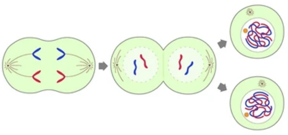
Telophase: Two New Dance Troupes Form
Two things happen in telophase. First, fragments of the nuclear membrane broken down at the end of prophase are recycled to create two new nuclear membranes around the chromosomes in the daughter cells. Second, the cell pinches inward at the equator. Cell parts (e.g., organelles, macromolecules, metabolites and ions) will be equally distributed around the parent cell such that when the cell does pinch off (a process called cytokinesis) each will have what it needs to function. Finally, the compacted chromosomes de-compact in their new nuclei. This completes cell division.


Comments