5. Tiny Machines (1,111)
- lscole
- Apr 11, 2025
- 5 min read
Updated: 15 hours ago
In the last chapter, we described a system that seems almost paradoxical: a cell filled with molecules moving randomly--colliding, binding, and separating--and yet somehow producing precise, coordinated behaviors. The question is: what makes that possible? What are the molecules that take these countless random interactions and turn them into consistent, functional outcomes?
The answer is not a central controller. The answer is proteins.
A typical human cell contains tens of millions of protein molecules. Some cut DNA. Others build new molecules, transport cargo, or transmit signals. Some act as motors, pumps, switches, and clamps--literally tiny machines carrying out the work that keeps the cell alive.
But what makes proteins remarkable is not just what they do. It’s how they do it.
Proteins don’t plan, and they don’t know what they are doing. They are simply molecules, subject to the same random motion as everything else in the cell. And yet, when they collide with the right partners in the right orientations, they reliably produce specific outcomes.
To get some perspective, proteins account for about 50% of the cell's dry weight. That's much more than DNA, which accounts for about 1-2%. It's also more than RNA, which accounts for about 20%.
But it isn't just their quantity. As I described, proteins are the workhorses of the cell. RNAs can play functional roles, too. But even when RNAs act directly, they usually do so as part of protein-RNA machines. To keep our model of the cell simpler, we'll focus only on proteins.
Many different proteins
There are two broad categories of proteins based on their role. Functional proteins perform tasks and tend to be globular, or somewhat spherical in shape. Structural proteins provide physical support to the cell and often take the form of elongated fibers. But there are exceptions.

The variety of different kinds of proteins in the cell is staggering. A human cell can produce tens of thousands of different proteins, each folded into its own unique shape with its own unique task.
Much of protein diversity is due to the many kinds of functional proteins. There are many more different kinds of functional proteins than structural proteins.
Functional protein roles
Functional proteins will be the focus of this book--more so than structural proteins. Many fall into larger categories of which there are many kinds. Enzymes speed up chemical reactions. Motor proteins carry cargo from one part of the cell to another. Transcription factors turn genes on and off. Nucleases cut DNA during the process of DNA repair. Signaling proteins attach to receptors on other cells enabling cell-to-cell communication.
Different proteins do different jobs--but most operate in a similar way: by binding, changing shape, and releasing. It is the shape change (or "conformational change") that does the work. This is how proteins work. Next let's look at what a protein actually is.
A chain of amino acids
How does the cell create thousands of completely different protein machines efficiently? By using a limited set of subcomponents and mixing and matching them liberally.
In fact, proteins are a general kind of molecule called a polymer. Polymers are long molecules made of repeating subunits. Proteins are polymers. So is DNA.
Only 20 standard amino acids--the only subcomponents of proteins--are needed to build every protein in the human body. These 20 amino acids are chemically different from each other, but similar in that they're all capable of attaching to each other in linear manner. On average, a human protein might be composed of 300-400 amino acids all lined up and connected. But they can range from 50 to thousands of amino acids long.
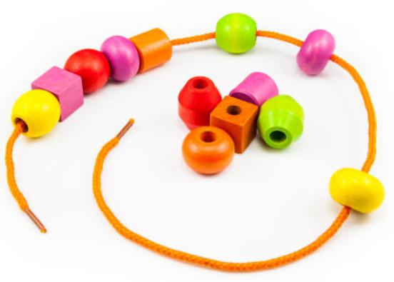
Imagine 20 different colored beads threaded onto a string. Any number of beads can be on the string. They can be in any order.
Each of the differently colored beads represents a different amino acid. We'll call the protein's linear order of amino acids its linear structure.
From chains to tiny machines
A newly made protein chain doesn’t stay linear for long. Within milliseconds the chain twists, bends, and snaps into a precise three-dimensional shape.
This automatic folding occurs because different amino acids have different chemical properties. Some are positively charged; others are negatively charged. Some are large; others are small. Some are hydrophilic (attracted to water); others are hydrophobic (repelled by water).
A protein’s final shape is not imposed--it emerges from countless local interactions between its parts. Each amino acid responds to other amino acids in its local environment differently.
Protein folding, by the way, can also be viewed as an instance of the general concept of emergence. No one directs protein folding. It arises from the chemical properties of the component amino acids. Folding is based on nothing but blind interactions between the different amino acids in the chain. But those myriad individual local interactions result in a perfectly folded functional protein.
We'll call the protein's folded form its folded structure. This is the functional three-dimensional form of the protein--the form capable of doing the kinds of jobs I've described. And doing these jobs almost always involves the protein changing its shape.
In other words, proteins don’t act simply by attaching to other molecules. Binding often triggers a change in shape--a conformational shift that alters what the protein can do next. These changes often occur in cycles: binding, shifting shape, carrying out a function, releasing, and returning to the original state. In this way, proteins operate less like static tools and more like dynamic machines, cycling through different states as they interact with their surroundings.

Multi-protein complexes
Finally, in some cases, multiple folded protein structures self-assemble to form a multi-protein complex. Multi-protein complexes can contain a few proteins or dozens. These are referred to as "dimers," "trimers," "tetramers," and "pentamers," etc. The proteins can be the same or different. When they're the same, we use the prefix "homo-". When they're different, we use the prefix "hetero-".
In many cases, proteins self-assemble into these larger machines—structures that, like individual proteins, arise from local interactions rather than central design. This, by the way, is the same kind of emergence we saw earlier between amino acids. In this case it's local, blind interactions between individual proteins resulting in a new, functional protein complex. We'll be introduced to many multi-protein complexes and individual proteins once we get into genome replication.
To summarize, proteins are where random motion becomes directed activity. Proteins don’t plan and they don’t know what they are doing. But when they collide with the right partners in the right orientations, their shapes change in ways that produce consistent, tangible results. From these countless local interactions—binding, changing shape, releasing—coordinated cellular behavior emerges.
In the next chapter, we'll get to know DNA.

Comments